Automated Analysis Of Protein Secondary Structure Changes Due To Ligand Binding Using Microfluidic Modulation Spectroscopy
Webinar Length: 1 hour
In this webinar you will learn:
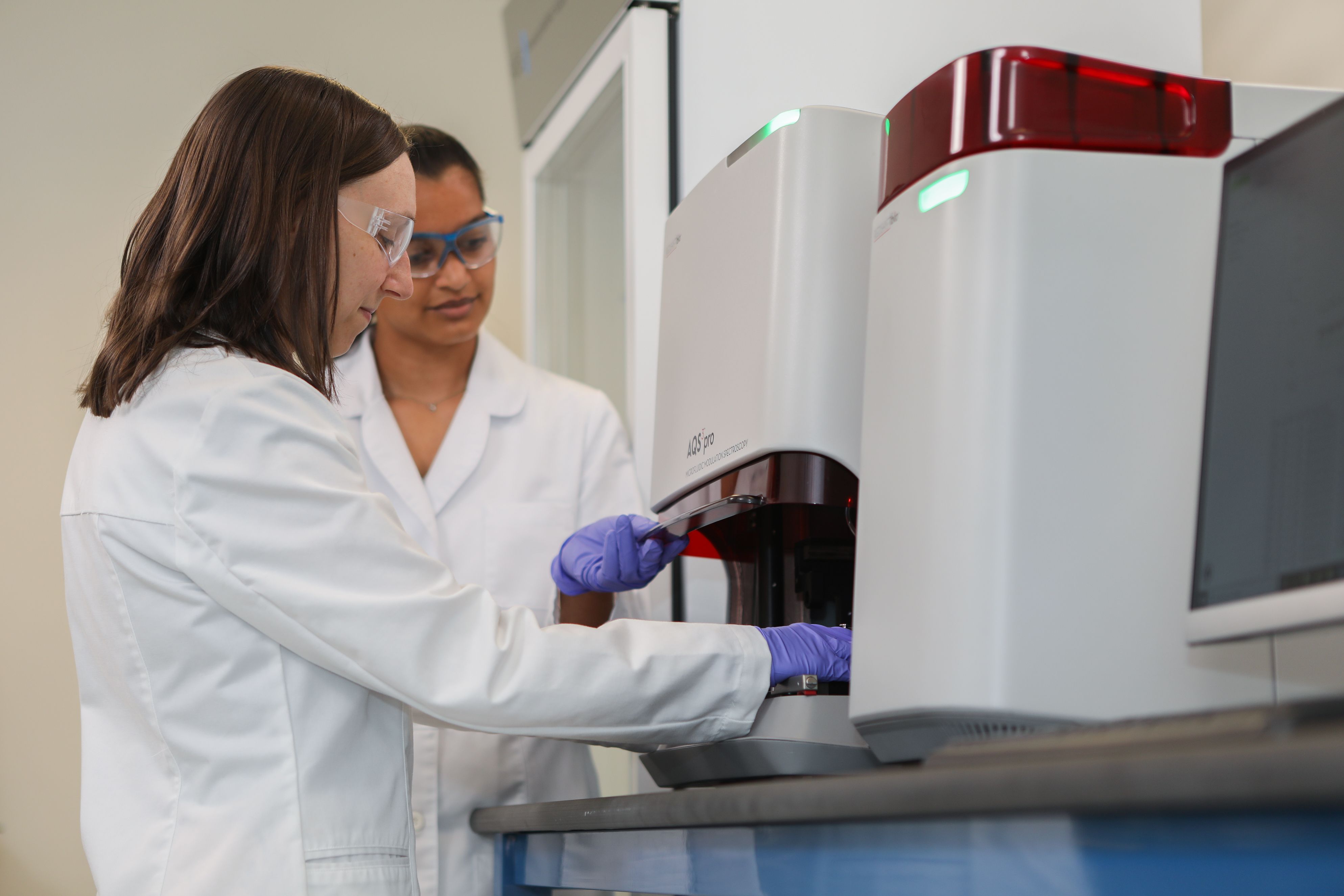
Ligand binding can affect the function of proteins and often causes conformational change in the protein target. Since form fits function, determining the secondary structure of proteins with and without ligands is essential for a more complete understanding of how the ligand alters the protein’s structure and function. In this study the AQS3pro, developed by RedShiftBio, was used to measure protein secondary structure of 2 different ligands bound to their substrate protein. The AQS3pro, featuring Microfluidic Modulation Spectroscopy (MMS), measures protein structure by combining infrared spectroscopy with microfluidics to enhance the sensitivity and accuracy of IR spectroscopy. Using a Quantum Cascade Laser (QCL) that is 100 times brighter than FTIR light sources, MMS has enabled the ability to probe secondary structure more precisely and over a large concentration range (0.1-200+ mg/mL), all while using fully automated sample handling to minimize instrument hands-on time and maximize sample throughput. Additionally, MMS uses a flow cell that modulates between sample and reference buffer, using real-time, automated buffer subtraction that is highly accurate and compatible for use with complex buffer systems including organic modifiers such as DMSO.
MMS was used to investigate the stabilizing effect of two ligands on a protein substrate. In the data shown, the first ligand provides a strong stabilizing effect, whilst the second ligand provides a partial stabilization effect when the samples have been exposed to higher temperatures. This data provides insights into the binding properties of the two ligands. The webinar demonstrates that overall, MMS is a sensitive secondary structure characterization tool that can enhance the biophysical characterization toolbox by contributing secondary structural information to ligand binding applications.